Neuro-Cardiac-Guided Transcranial Magnetic Stimulation (NCG TMS)
Targetting the DLPFC: Use of Target Engagement
Using rTMS in the treatment of depression, clinicians can rely on various stimulation targets. Overall the consensus is that rTMS should target the Dorsolateral Prefrontal Cortex (DLPFC), however there are several heuristics to apply this in practice, such as the use of the 5-cm. rule, the Beam-F3 method or structural MRI Neuronavigation. While all three methods aim to target the same structure, within the same individual, these heuristics could result in substantial discrepancies. On the other hand, clinical efffectiveness of these measures is rather comparable on the group level, demonstrating remission rates between 30-37% (Blumberger et al., 2019; Carpenter et al., 2012; Fitzgerald et al., 2016). Therefore, it would be valuable to have a technique available to estabish ‘target engagement’ by which one can verify that the right network is targetted, rather then relying on a heuristic based on ‘assumptions’.
Target engagement comprises the use of a direct functional outcome measure as a validation for targeting the correct TMS location, whereby it can be demonstrated that the right area is activated, either directly or trans-synaptically. In the same way as the motor cortex is identified by thumb movement as a demonstration of primary motor cortex activation, such functional outcome measures are thus far lacking for the prefrontal cortex and DLPFC. One proposed method is by extracting connectivity patterns to frontal areas using the sgACC as a seed region (Fox et al., 2012; Fox, Liu, & Pascual-Leone, 2013). However, such approaches require an fMRI scan from every individual patient, which is less practical in clinical practice, but above all this method has shown poor interindividual reproducibility (Ning et al., 2018), limiting its use on the individual level.
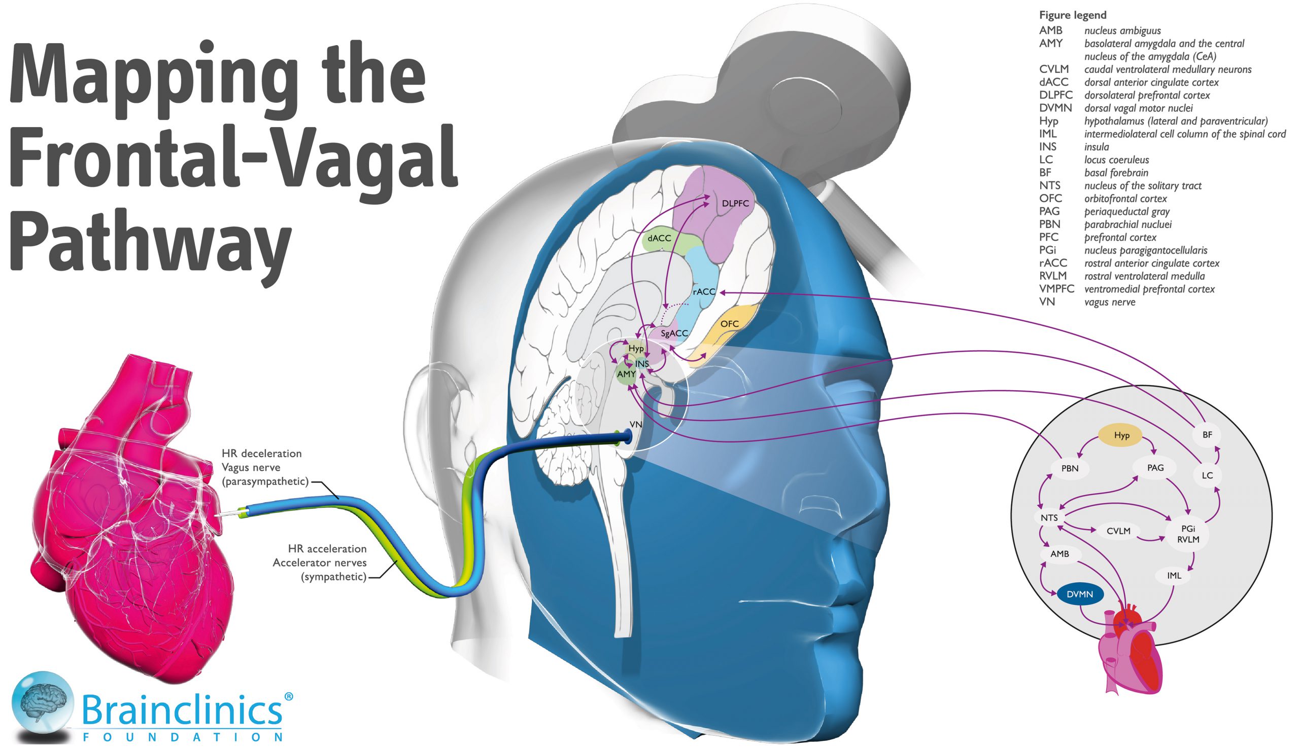
In 2017, we published a first pilot study on a novel method that employs heart rate assessed during TMS stimuation, to verify ‘target engageent’ of the Frontal-Vagal network, that is hypothesized to be involved in Depression (see figure on the right and review from Iseger et al., 2019 in the PDF reader below) which we termed Neuro-Cardiac Guided TMS or NCG TMS.

NCG TMS: How it works.
The depression network and the brain-heart axis are interconnected and stimulation of the DLPFC was shown to reduce heart rate (Makovac et al., 2016). The parasympathetic effects on the heart are short-lived; stimulation of the vagus nerve therefore usually results in an immediate response of the heart, typically occurring within the cardiac cycle in which the stimulation occurred, with a peak in heart rate deceleration within 5 seconds (Buschman et al., 2006). The return to a normal HR is very quick after the activity of the vagus nerve is reduced (Shaffer, McCraty, & Zerr, 2014). Also see the illustrated movie on the left for a visualization of this Heart-Brain Coupling method and how it can inform if the 5 cm. or the Beam-F3/4 method is the right target for activating the Frontal-Vagal pathway.
In a recent study, it was shown that stimulating different prefrontal scalp locations led to different effects on heart rate, and that stimulation of the F4 and F3 locations (based on the 10-20 system) resulted in the most significant heart rate decelerations, whereas this was not found for central sites overlying the primary motor cortex or the posterior site Pz (Iseger et al., 2017). Individual variation was also demonstrated, however (i.e. for some individuals the most profound heart rate deceleration was found for slightly more posterior sites e.g. FC3, FC4 relative to F3 and F4), indicating that the Heart-Brain Coupling method could be used to individualize the correct stimulation target, under the assumption that trans-synaptic activation of the sgACC indeed activates the whole Frontal-Vagal pathway that is involved in the pathophysiology of MDD. Since this early observation in 2017, this finding has been independently replicated by Kaur and colleagues (2020) as well as in our own lab. See the review and first individual participant meta-analysis synthesizing all these data in the Iseger et al. review below in the PDF reader.
Further reading
The Heart-Brain Coupling app for iOS
The Brainclinics Foundation developed Heart-Brain Connect, a breakthrough Heart-Brain Coupling app to assist clinicians in pinpointing the optimal location for rTMS treatment. Our unique approach has great advantages over more traditional methods and can be tried for free. The app can also be used as a resonant breathing training method.
For more information, please visit this page.
Heart-Brain Connect is available in the iOS App store.
Free ebook
Brainclinics and Utrecht University alumna Tabitha Iseger’s excellent PhD thesis specifically on this topic of the Frontal-Vagal pathway in Depression and Heart-Brain Coupling can be downloaded free of charge from our website (as a pdf) or from Apple’s Bookstore.
We can further recommend some extra fine reading material if you’re interested in our publications, and on the right hand side you can browse the recent review and meta-analysis by Tabitha Iseger and colleagues, that also includes all references from the text above.
